Our main research areas are listed on this page.
To explore datasets from our neuroscience experiments see our
Brain Viewer.
To download our MRI toolbox for accelerated imaging please go to MRITools.
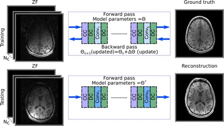
Compressive sensing (CS) is a powerful framework for reconstructing accelerated MR acquisitions, yet not without limitations. CS assumes sparsity priors in known linear transform domains (e.g. Wavelet, finite differences) to enable recovery, but these assumptions are often suboptimal. Since CS formulations weigh sparsity priors against experimental evidence (i.e., acquired data), careful hyperparameter tuning is essential to reconstruction performance. Lastly, image reconstruction is attained via nonlinear optimization algorithms with heavy computational burden.
We are currently developing novel MRI reconstruction algorithms based on deep neural network (NN) architectures to overcome limitations typically associated with CS. In the NN framework, prior information specific to MRI data is learned off-line from a large collection of MR images. For applications where data are relatively scarce, domain transfer can be performed to enhance learning, even between natural and MR images. Deep architectures with millions of free parameters are then employed for recovery that are only weakly sensitive to hyperparameters. Once trained, NN reconstructions can be performed online on the order of hundreds of milliseconds.
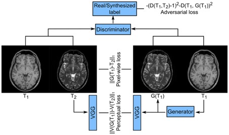
MRI offers unparalleled and flexible contrast generation mechanisms, which enables the same anatomy to be imaged under an array of unique tissue contrasts via tailored pulse sequences. For instance, the brain can be imaged under T1-weighted contrast to delineate grey and white matter, and under T2-weighed contrast to delineate lesions and fluids from heathy tissue. Although multi-contrast MRI provides broadened information to help improve diagnostic accuracy, prolonged scan times, incorporative patients and associated costs often render it impractical.
The ability to recollect completely missing or heavily corrupted images in a multi-contrast MRI protocol can greatly improve clinical practice. Towards this aim, we are devising novel methods for image synthesis and reconstruction in multi-contrast MRI based on generative adversarial networks (GAN). These architectures involve a minimax game play between a generator that is geared to recover realistic images of desired contrasts given collected source contrasts, and a discriminator that aims to separate real MR images from those synthesized by the generator. The resulting GAN models are much better at preserving textural details than traditional methods, and they enable use of paired and unpaired datasets.
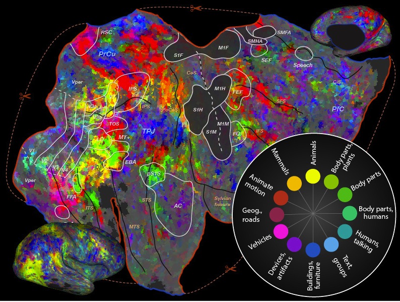
Previous studies of visual category representation have identified a limited number of regions in the human brain that are highly responsive for specific categories. Well-known examples of these specialized regions include the fusiform face area (for faces) and the extrastriate body area (for bodies). These results have led to the hypothesis that category representation is mediated by highly localized and dedicated regions. However, humans can recognize thousands of unique visual object and action categories in natural scenes. It is therefore unlikely that a distinct brain region can be dedicated to each category.
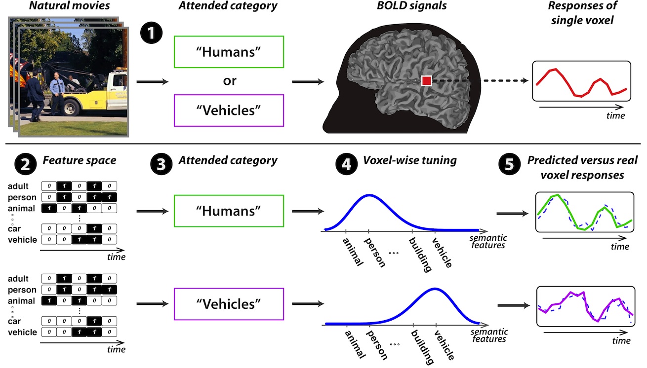
To reveal the details of category representation across the entire brain, we are using natural movie stimuli that contain thousands of object and action categories. We first record brain activity with functional MRI while participants passively view these movies. We then employ a powerful voxel-wise modeling framework to quantitatively describe the selectivity of each voxel to each category. Our results imply that many brain regions represent information about a broader set of categories than previously assumed. We are currently examining the details of category representation across both visual and non-visual brain regions.
Although real-world scenes are cluttered with many different objects, humans are extremely adept at finding target objects in natural environments and shifting their attention rapidly between different targets. The precise neural mechanisms that mediate this remarkable ability are yet unknown. Previous studies have reported relatively simple mechanisms that increase the quality of brain activity evoked by attended objects, without affecting the way that information is represented in each brain region. Yet, because there are a limited number of cortical neurons, it seems unlikely that all brain regions retain fixed representations irrespective of behavioral demands.
To study the nature of neural representations during visual search, we use complex natural movies and a powerful computational modeling technique to describe the relationship between visual information and brain activity. Our preliminary results suggest a novel attentional mechanism that reallocates neural resources across the brain to maximize sensitivity for the target and improve target detection under demanding conditions. You can explore our datasets here. We are currently studying the generality and flexibility of this mechanism for vision and for other sensory modalities.
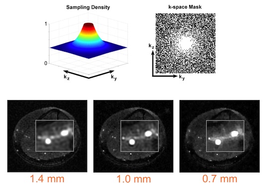
Recently developed compressed sensing (CS) theory has opened up great avenues for accelerating MRI exams. The CS theory foresees that a compressible image can be sensed from a significantly smaller number of data samples than the Nyquist rate. To recover an image from these reduced set of samples, a linear transform of the image should be well represented by few coefficients. Furthermore, missing data samples should introduce aliasing artifacts with noise-like incoherent structure. MRI images exhibit similar statistics to natural images, and they are often highly compressible. However, the optimal sparsifying transforms and data sampling patterns for distinct body regions are not fully known.
We have ongoing efforts to apply CS to various MRI applications including MR angiography, MRI stem-cell tracking, and neuroimaging. We are developing novel automated parameter tuning algorithms that find dataset-specific reconstruction parameters to maximize image quality. These algorithms will empower practical use of CS reconstructions in clinical environments where manual tuning is impossible.
MRI is a powerful modality that depicts the morphology and function of biological tissues noninvasively. Despite its increasingly important role in both clinical and research environments, image quality and spatiotemporal resolution in many MRI applications are limited by prohibitively long scan times. Thus, there has been a growing need for accelerated MRI methods.
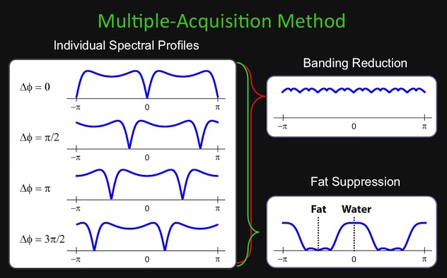
To overcome the speed limitations of conventional MRI, we are developing a class of rapid pulse sequences based on steady-state free-precession (SSFP) imaging. SSFP sequences offer high signal-to-noise ratios, but can yield suboptimal tissue contrast and image artifacts due to signal loss. We tailor intelligent reconstruction algorithms to improve tissue contrast and artifact suppression in SSFP images to clinical standards.
